Firearms of suspicion, questioned bullets or casings, cartridges are examined so that the mysteries it holds can be resolved down to certainties and some conclusion. These examinations are accomplished with skilled hands, thorough knowledge and with precautions. This examination of firearm can be of various types depending upon the requirement and the interest of the question to be solved.
MICROSCOPIC EXAMINATION OF FIREARM
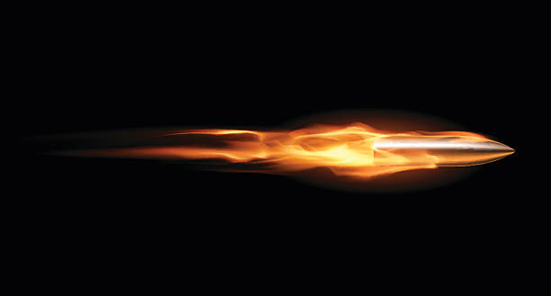
Examination of firearm or its parts (bullet, cartridge etc.) holds high evidential value of only they can be examined through skilled hands and intellectuality. There are several phenomena and process behind working of firearm and hitting any target. Bullet especially bears several marks which if seen under microscope may reveal many information. It may even lead to the tool it has been shot with. Utilizing the efficiency of expert and microscope various facts can be established and can be compared to suspected firearm for the proof.
Stereomicroscopy
Stereo microscope provides the best and clearer output when bullets are subjected to comparison. They provide three-dimensional and this is why examination through stereoscopy overrules all other methods. Stereoscopy can also deal with the identification of the cartridge cases with supreme and positive result. Through Stereoscopy one can understand the direction of investigating comparison between bullets through comparison microscope Surfaces of the exhibit can be tilted and rotated at any angle to provide better result.
Comparison Microscope
When it comes in compare the ballets if they were shot from the same firearm or not, nothing can works like the comparison microscope. It is the amalgamation of two individual microscopes in which two objective from both the microscopes forms the single image by adjusting focus in the same eye piece through prism. The whole set-up is connected to computer for better visualization of every single minutiae and to save the data by directly clicking the image through computer and saving it for future use. The arrangement also has mechanical stages of process by moving exhibits up, down, left and right and also by adjusting focus for feasible intensity of light and exposure.
Some of these arrangements comes up with fluorescent lamp system either with single tube-light or double tube-light. The use of comparison microscope is extremely vital in forensic science as it contributes matching of the most evidential marks and features in ballistics like breech scrape marks, striations on primer even those formed due to interference of cartridge, partial sliding marks in the barrel, breech face marks.
Firing pin marks by evaluating its periphery to examine multi-firing, marks in the chamber like marks that are formed due to joint of the bore in the chamber, marks of the ejector and extractor that are formed on the cartridge due to low-pressure and also marks on the bullet that are formed in the smooth bore barrel. Sometimes there are certain designing or specific marks are generated in the firearms while their manufacturing, these aids as individual marks and are accessible to examine for the comparison.
Chemical Examination
Examination of firearms and its effects caused through chemical properties is a next step in the evaluation. In this process we observe the presence of exhibits in the samples which are under suspicions. Certain chemical and combination of chemical is brought together in such a way that it should give a confirmatory or at least indicative result either in the form of precipitate or through some changes in the color of the final product. Following are few most relevant test regarding evaluation of ballistic exhibits.
Sodium Rhodizonate Test (Price’s Spot Test)
It is one of the most important test in ballistics as it is used for testing the presence of lead in the sample. This procedure is very feasible and inexpensive especially when microscopic examination for determining the presence of lead is unclear. In this process GSR is treated with 1% HCI, then the sample is dries with hot air with spotting of sodium rhodizonate in water. Sample will now tum into orange color.
Now again apply gentle warmth and again treat it with solution of 0.1 N HCI but do not dry it further. Orange color will start disappearing but if there are traces of GSR it will stay there in purple coloration. For more positivity is the result the last product can be treated with barium and gives the brown color.
Walker Test for Nitrites
This test is done to check the presence of nitrites in the sample as nitrites are the components found in unburnt as well as in partially burnt propellant. The test is conducted by using gelatinous surface of desensitized photographic paper so as to collect sample from cloth or from other fiber as they are quite sticky and unburnt or partially bunt particle can easily adhere to this paper.
Now convert these into a diazo dye compounds cany color like bright orange color for clear visibility). Then numb the sheet of photographic paper by using hypo fixer. Now dip it into a solution of 5% 2-naphthylamine-4,8-disulphonic acid and then air dry the sample. Now treat it with 20% acetic acid an apply warm by ironing. If the bright spots in the red appears then the presence of nitrites is confirmed else it’s a false sample.
Marshall Test
After collecting sample through desensitized photographic paper treat it with0.5% solution sulphanilic acid for 10 min Now soak the same sample in a 0.5% solution of N-naphthyl ethylenediamine hydrochloride in methanol for 2 minutes and then again air dry it. The treat it with 20% acetic acid after which apply pressure with warm iron for five minutes. Spots of purple color will appear on the purple backgrounds for better visibility rinse the sample with warm water and the background color will be easily eliminated without hampering the developed positive spots. Now rinse these spots with methanol and notice the change in color from purple to orange for the confirmation.
Tewari Test
Dissolution of antazoline hydrochloride (2 N-benzylanilinomethyliminazoline hydrochloride) in 50 ml. of water. Now add 45 mL. of conc: HCI and blend it until the white precipitate runs out of visibility. Now immerse a filter paper in acetone and apply it on target. Now, air dry the sample and then and spray severely with the prepared antazoline solution. Deep yellow spots will appear and confirms the presence of nitrite compound.
Lange Reagent
This test is also known as Dermal Nitrite Test. It is used to detect whether the firearm was used or not. 0.25% solution of diphenybenzidine in concentrated Sulphuric acid is added. Now spray this reagent onto paraffin casts of the suspect’s hands. Deep blue color will appear if there will be any trace of nitrite particles that involves nitrocellulose. Further in later use Diphenybenzidine was replaced with diphenyamine as they were very carcinogenic in nature. This amendment gives the same result that is deep blue coloration to nitrites. But since test gives coloration for every other sample containing nitrite like-fertilizer hence the process was demolished as it was unreliable.
Harrison and Gilroy Reagent
Like as in other test collection will be again from photographic paper to lift the reside the only change here will be to use dilute HCI rather using acetic acid. After air dry spray 10% solution of triphenylmethylarisonium iodide in alcohol over the sample. Orange coloration will appear which is confirms the presence of antimony.
But if after air dry sample is sprayed with a saturated solution of sodium Rhodizonate, red spots will appear and confirms the traces of barium or lead. Similarly, if the chemical is changed again and is sprayed with dilute HCL After air drying purple spots will appear for positive result for lead. Similarly red coloration will appear if after air dry, the sample is exposed to a 35% ammonia solution and confirms the presence of barium.
Instrumental Examination
Concurrent viewing of the magnified images of every detail is possible through comparison microscope but in earlier times, through normal microscope study of minutiae was not possible on a satisfactory level. With time and evolution, revolution in technology has now changed the
working process and skills of the experts as well. In comparison microscope the images of exhibit are parted through a fine line through the center of the field. This aids in through study and storage of exhibit at the same time. Besides microscope or comparison microscope there are some other techniques which delivers plenty of other detailed information hidden in the sample unconventionally. These are discussed as below:
Rifling Mater
This instrument is used to measure the actual twisting of rifle. To process, clamping of the barrel is done to the lathe bed and in the tail of the stalk, it is fixed from its one end by a long steel rod which has lead of same diameter as the bore bears while the other end is attached to a progressed disc that rotates accordingly. The rod from the one end is pushed causing the firearm to turn as rifling nibbles into the plug. The distance that the rod travels down the bore estimates the grade of gyration. Although the measuring of rifling is barely faced by experts of the discipline.
Comparison Camera
Essentially, a comparison camera is a camera with an exceptionally extended frame with two lenses. Here are two stages in front of the lens on which the exhibits can be placed and can be evaluated. These stages are operated with stretchable rods to adjust exhibits to the maximum evaluation. Over in all it simply aids to compare exhibits side by side with a higher range of magnification although this technique is rarely used by experts in contemporary cases.
Neutron Activation Analysis (NAA)
It is highly sensitive technique although it time consuming and is not at all feasible for detection and evaluation of lead which the most important element of GSR Through NAA. chiefly evaluation for GSR is done. This examination is based on the presence of antimony and barium. When the sample is exposed to NAA and if the antimony or barium is present in it, it will absorb on neutron and will release a radioactive particle of antimony or barium. These released products are comprised of gamma rays and can be evaluated for isotopes by calculating half-life by comparing it to standards
Atomic Absorption Spectrometry (AAS)
It is the most convenient, sensitive and feasible technique for evaluation of GSR and can detect traces of element of interest even in nano and Pico grams. It is cost friendly technique which consume much less time than any other technique. Under this process the sample is exposed to wavelength of the same as of the sample emits when it’s in excited position. So the GSR collected is taken into graphite tube and is heated for excitation. Now the discharged radiation from the sample is passed through atomizer. The intensity is measured for any changes in quantity of the element of interest.
Scanning Electron Microscope (SEM)
This technique is very remarkable and efficient for the evaluation of GSR. The technique noninvasive in nature and delivers the result data in particles per unit area. Its accuracy and competency is way out of question and hence is very reliable technique in the ballistics. Under this process the sample is exposed to the beam of electron. This electron beam is having certain impacts as it produces X-rays, luminescence effects, back scatter and delivers maximum resolution power. This technique is quite obligatory regarding examination of ballistics.

X-ray fluorescence
All the methods and techniques for evaluating GSR has been discussed above but there is one more methodology through which GSR can be examined well which is X-ray fluorescence. This method is primarily used for quantitative and qualitative output from the sample of interest. It is considered the most convenient method of all because of its simple handling and operating methods which gives the result of highest accuracy and is best if the sample is in the powdered form.
For more updates, subscribe to our blog.