

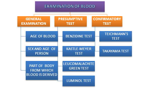
The number of techniques are used in the examination of blood. They involve physical, presumptive and confirmatory examination of blood.
GENERAL EXAMINATION OF BLOOD :
- Age of blood: On examination of blood fresh stains on light colored clothes are of bright-red color, which gradually changes to reddish – brown in 24 hrs. and brown within few days after this blood becomes black. Fresh blood is moist and on drying it stiffens because of blood. In an ordinary condition, the bloods usually dry within an hour or two. The recently shed arterial blood is bright red and venous blood dark-red.
- Sex and age of person : By examination of blood sex can be determined by the presence of sex chromatin in the leucocytes, if the cells can be identified. The presence of foetal haemoglobin indicated that blood is derived from a child.
- Part of body from which blood is derived: menstrual blood is usually found on female garments, diaper or piece of cloth. It is a dark and fluid reaction is acidic and contains fibrinolysins and elevated levels of LDH4 and LDH5. modernforensic.in
CHEMICAL EXAMINATION OF BLOOD:
PRESUMPTIVE TEST: It is also known as color test. It the questioned sample gives positive color test in any two color test, the sample is possibly the blood. If it fails to give color reactions, in all probability it is not a blood sample. In presumptive examination of blood 4 tests are included.
- BENZIDINE TEST/ TETRA METHYL BENZIDINE TEST:
Reagent:
- TMB solution 1.5g
- Glacial acetate 20ml
- 3% Hydrogen peroxide
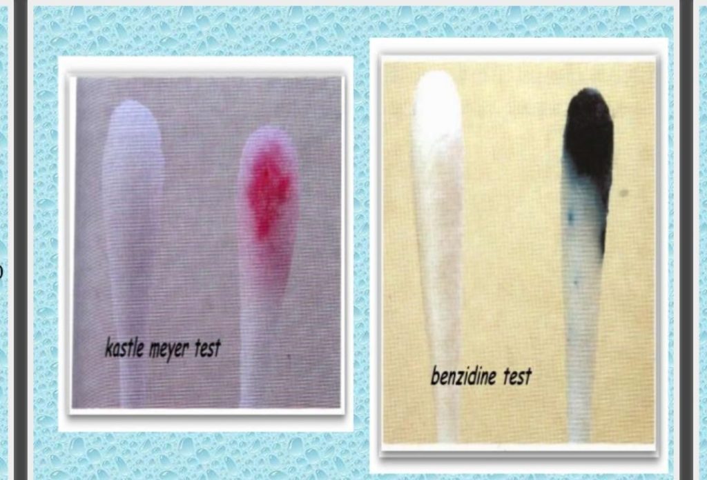
Take a blood sample and place it in a porcelain tile. Add some drop of saturated benzidine solution in glacial acetic acid and then a drop of 10 volume hydrogen peroxide. If the blood is present, dark blue color is produced immediately.
This is the best presumptive test for blood as it detects blood when present in dilution of one part of in three lakhs. The positive reaction is not a proof that the sample is blood, but negative reaction rules out of the presence of blood.A positive reaction is given by blood of almost any age, blood that has been exposed to heat and treated with cleaning agent.
2. PHENOLPHTHALEIN TEST (KASTLE-MEYER TEST):
Reagent :
- Phenolphthalein solution 2ml
- Hydrogen peroxide 10ml
In a test tube containing blood sample, add20-40 drops of phenolphthalein reagent (phenolphthalein 2g+ sodium hydroxide 20gm +zinc+ distilled water) and then a drop of hydrogen peroxide. If the blood is present, a pink or purple color immediately appears. This test is more specific than benzidine but less specific.
3. Leucomalachite green test:
Reagent:
- Leucomalachite green 0.1 g
- Sodium perborate 3.2g
- Glacial acetic acid 66ml
- Distilled water 33ml
Take a blood sample and add leucomalachite green solution and sodium perborate in 65% glacial acetic acid and distilled water . if green color appears it indicates the presence of bloo
4. Luminol test:
- Luminol reagent :
- Sodium perborate 0.7g
- 3-aminophthal hydrazide 0.1g
- Sodium bicarbonate 5g
In this test take the blood extract and add few drops of luminal reagent ( sodium perborate and 3-aminophthal hydrazide in sodium bicarbonate). Appearance of fluorescene color indicates the positive test for blood.
CONFIRMATORY TEST FOR BLOOD
This basic principle of chemical examination of blood is based on the property of heme (iron) part of haemoglobin to form characteristics colored crystals with certain reagents. in confirmatory examination of blood 2 crystals tests are involved.
- HAEMIN CRYSTAL TEST/ TEICHMANN’S TEST:
Teichmann’s reagent :
- KCl, KBr and KI 0.1 g each
- Glacial acetic acid 100ml
Add some drops of solution of potassium iodide, potassium bromide and potassium chloride to the slide of blood sample . A cover slip is placed and on warming, it gives out bubbles on addition of a drop of hydrogen peroxide if it is blood. It is examined under microscope after cooling. Brown-black rhombic crystals of heamin or haematin chloride are obtained in clusters.
The reaction is negative if the stain is old, is washed or treated by chemicals, over-heating.

2. HAEMOCHROMOGEN CRYSTALS TEST/ TAKAYAMA TEST: the takayama test uses takayama reagents which is a solution of following:
- Saturated glucose solution (10%)
- potassium hydroxide solution (10%)
- Pyridine solution and
- Glacial acetic acid
On warming, the dry blood crust with the takayama reagent gives needle shaped crystals of hemochromogen. The main reactions involved are:
sodium hydroxide releases the heme from the globin through alkaline hydrolysis. Glucose releases the heme iron and Pyridine combines with it to form the product, pyridine ferroprotoporphyrin.
SPECTROSCOPIC EXAMINATION OF BLOOD :It is the most delicate and reliable test for detecting the presence of blood in recent and old stains. Less than 0.1 mg of blood is sufficient.
The blood haemoglobin is changed in 2-3 forms on the slide itself and characteristics absorption is observed. Usually alkali haematein (obtained by treating the stains with ammonium sulphide) and cyanhaemochromogen (obtained by treating the haematein with potassium cyanide) are studied for absorption spectroscopy.
EXAMINATION OF BLOOD SPECIES
This examination of blood species determines whether the blood is from human being or from a lower animal.
- Precipitin test: blood serum contains proteins in a colloidal and a suspension, and when human serum is injected into an animal, the animal becomes immunized against these proteins and anti-bodies develop inn its blood. If human serum is then brought into contact with this animal serum, then antibodies in the animal serum reacts with the proteins in the human serum and a visible precipitate forms. The antibodies causing this reactions are known as precipitin serum. A suitable antiserum should react immediately or within a minute on the 1:1000 dilutions.
Procedure: the presence of blood is determined first. An extract of stained material is prepared by soaking it in normal saline. The extract should be clear and may be filtered or centrifuged if necessary. Two drops of undiluted antiserum is added to the extract in a small test tube. At the junction of two fluids a white ring appears in the case of positive reaction. In case of negative reaction, no ring appears. The results are qualitative and expressed as positive or negative.
2. Double-diffusion test: in this test both the reactants, antigen and antibody diffuses towards each other in agar gel plate and when an antigen combines with its specific antibody at opium proportions, precipitin are formed. In this method fill the central well with the tissue extract and peripheral wells with different anti-sera for species origin like ( anti-human serum, anti-dog serum, anti-cow serum etc.). Cover the petri dish and keep it overnight. Examine the gel for the presence of precipitin hand form. The advantage of this method is that the extract can be tested against different anti-sera simultaneously.
2. Crossover electrophoresis: the stain extract (antigen) is placed in the cathodic well and the anti-serum in the anodic end. When electric current is applied the antibodies migrate catholically. The other serum proteins migrate anodically. A precipitin reaction occur midway between the paired well, when an antigen combines with its specific antibody.
Fore more such blogs, subscribe to our blog.